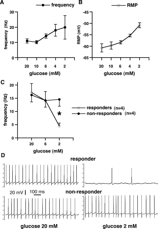
Figure 7.
Single-cell recordings in the GABAergic cells of the SNR during decreases of extracellular glucose. A, Whole cell, 2 mm ATP in the electrode: frequency of GABAergic cell discharges (y-axis) is plotted versus extracellular glucose concentration (x-axis). The frequency of discharges significantly increased with decreasing extracellular glucose, reaching the highest firing rate at 2 mm extracellular glucose concentration (36 mg/100 ml). This indicates a significant contribution of presynaptic KATP channels in the increased postsynaptic firing, as postsynaptic ATP was clamped. B, Whole cell, 2 mm ATP in the electrode: resting membrane potential (y-axis) is plotted versus extracellular glucose concentration (x-axis). Resting membrane potential of the recorded cells significantly decreased in the decreasing glucose concentration. This finding is consistent with increased firing frequency. Furthermore, this illustrates that, in this case, the postsynaptic membrane cannot be affected by changes in intracellular ATP (clamped at 2 mm). Therefore, if postsynaptic intracellular ATP is kept constant, the effects of presynaptic ATP deficiency prevail, which result in a decreased release of GABA (During et al., 1995) from multiple GABAergic terminals predominantly of striatal origin (Bevan et al., 1996) and, thus, in increased postsynaptic firing. C, Gramicidin perforated patch clamp: frequency of GABAergic cell discharges (y-axis) is plotted versus extracellular glucose concentration (x-axis). This recording arrangement evaluated contribution of both presynaptic and postsynaptic KATP channels as postsynaptic ATP was allowed to fluctuate according to the metabolic state. The population of cells divided into two subpopulations: responders, which significantly decreased their firing rate with the decrease of extracellular glucose compared with nonresponders, in which the firing rate remained almost constant even after 10 min in 2 mm glucose concentration (p < 0.05, t test). In the hippocampus, this concentration of extracellular glucose may completely shut down synaptic transmission (Kirchner et al., 2006). D, Gramicidin perforated patch clamp: recording from electrophysiologically identified GABAergic neurons. The first neuron was spontaneously active with action potential firing at ∼19 Hz in 20 mm extracellular glucose concentration (top left). After decrease of extracellular glucose concentration to 2 mm, the frequency of action potentials decreased to 2 Hz (top right). This neuron is referred to as responder. Another neuron was spontaneously firing action potentials at ∼18 Hz in 20 mm extracellular glucose (bottom left). After extracellular glucose decreased to 2 mm, the firing frequency was maintained (bottom right). This neuron is referred to as a nonresponder.

