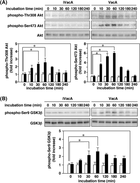
FIGURE 1.
VacA-induced Akt phosphorylation leading to GSK3β phosphorylation at Ser9. A, AZ-521 cells were incubated with 120 nm VacA or heat-inactivated VacA (iVacA) at 37 °C for the indicated times. After incubation, cell lysates were analyzed by SDS-PAGE (10% gels), followed by Western blot analyses using anti-phospho-Thr-308 Akt, phospho-Ser-473 Akt, and Akt antibodies. Results are representative of three independent experiments. Quantification of phospho-Thr-308 Akt and phospho-Ser-473 Akt obtained with VacA (filled bars) and iVacA (open bars) was performed by densitometry. Data are means ± S.E. of values from triplicate experiments, with an n = 3 per experiment. Statistical significance: *, p < 0.01. B, AZ-521 cells were incubated with 120 nm VacA (filled bars) or iVacA (open bars) at 37 °C for indicated times. Cell lysates were prepared at indicated incubation times and subjected to Western blot analyses using anti-phospho-Ser-9 GSK3β and GSK3β antibodies. Results are representative of three independent experiments. Quantification of phospho-Ser-9 GSK3β obtained with VacA (filled bars) and iVacA (open bars) was determined by densitometry. Data are means ± S.E. of values from triplicate experiments, with an n = 3 per experiment. Statistical significance: *, p < 0.05.

