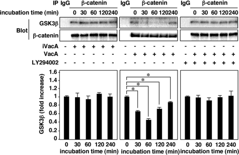
FIGURE 5.
GSK3β/β-catenin complexes are dissociated by VacA in AZ-521 cells. AZ-521 cells were incubated with 120 nm VacA or iVacA for 0–240 min in the presence or absence of 50 μm LY294002. After incubation, the cells were solubilized with lysis buffer and incubated on ice for 30 min. The lysates were available for immunoprecipitation using anti-β-catenin mouse monoclonal antibody (Transduction Laboratories, BD). The precipitates were subjected to SDS-PAGE in 8% gels and transferred to PVDF membranes. Then, GSK3β and β-catenin were quantified by Western blot analysis using anti-GSK3β rabbit monoclonal antibody (1:1,000; Cell Signaling) and anti-β-catenin rabbit polyclonal antibody (1:1,000; H-102, Santa Cruz Biotechnology), respectively. Quantification of phospho-Ser-9 GSK3β obtained with VacA (filled bars) and iVacA (open bars) was determined by densitometry. Data are means ± S.E. of values from triplicate experiments, with an n = 3 per experiment. Statistical significance: *, p < 0.01.

