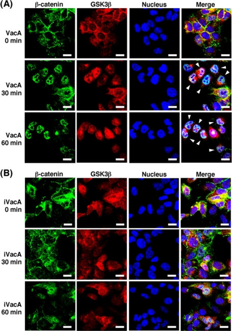
FIGURE 6.
Confocal microscopy analysis of β-catenin translocation to nucleus from cytosol. AZ-521 cells were incubated with 120 nm VacA (A) or iVacA (B) at 37 °C for 0, 30 and 60 min. After incubation, the cells were fixed using 2% paraformaldehyde at room temperature, treated with 0.1% Triton X-100, and blocked with Block Ace. The fixed cells were stained with DAPI for 5 min, and were subjected to triple immunostaining using anti-β-catenin monoclonal antibodies and anti-GSK3β monoclonal antibodies as primary antibodies. After treatment with the respective primary antibodies, cells were incubated with the secondary anti-mouse antibodies, conjugated with Alexa Fluor 488, and anti-rabbit antibodies conjugated with Alexa Fluor 546, respectively. Data are representative of three experiments. Scale bar, 5 μm.

