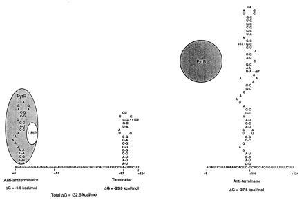
Figure 5.

A refined model for transcriptional attenuation of the B. subtilis pyr operon. The RNA sequences shown are from the first pyr attenuation (5′ leader) region. The RNA structure at the right is proposed to be the secondary structure allowing read-through and is the more stable structure when PyrR is not bound. The RNA structure at the left is proposed to favor transcription termination. The free energies of formation of the secondary structures shown do not assume any contribution from PyrR binding. The structure at the left is proposed to be stabilized by binding of the PyrR–UMP complex to the AAT stem-loop, which changes the free energy of formation to a value more negative than −32.6 kcal/mol (1 cal = 4.184 J). PyrR binds to pyr mRNA only when it is complexed with UMP.
