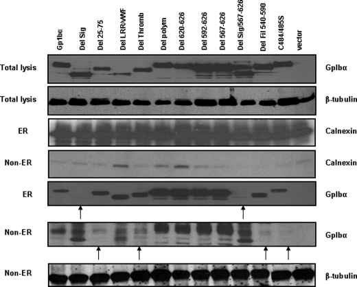
FIGURE 3.
Subcellular localization of mutant GpIbα proteins. ER-enriched and ER-depleted fractions from Rat1a cells expressing the GpIbα proteins depicted in Fig. 2A were subjected to Western blotting. Equivalent amounts of lysate from each fraction were electrophoresed. As a control for ER enrichment, the blot was probed with an antibody directed against calnexin and β-tubulin, which are ER-associated and non-ER-associated, respectively.

