
Figure 4.
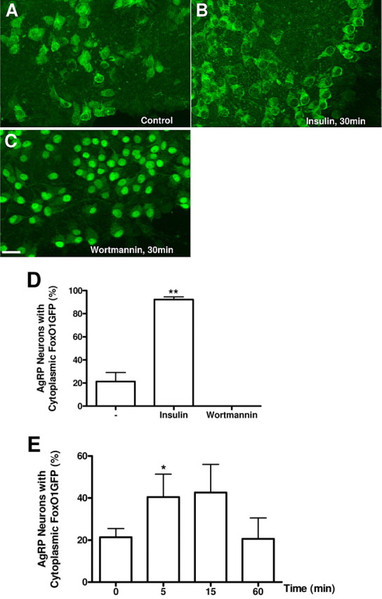
FoxO1GFP dynamics in AgRP neurons. A–C, Hypothalamic organotypic slices expressing FoxO1GFP in AgRP neurons were untreated (A) or treated with insulin (100 nm, 30 min) (B) or wortmannin (100 nm, 30 min) (C), and then fixed and stained with an anti-GFP antibody. Scale bar, 20 μm. D, Quantification of FoxO1GFP nuclear translocation in control, insulin or wortmannin treated organotypic slices of FoxO1GFP-AgRP mice. E, Effect of Leptin on FoxO1 subcellular localization in AgRP neurons. Slices expressing FoxO1GFP in AgRP neurons were treated with leptin (120 nm) for indicated time. Subcellular localization of FoxO1GFP is plotted as the percentage of neurons with cytoplasmic FoxO1GFP as in Figure 2. **p < 0.01, *p < 0.05 compared with control by unpaired Student's t test.