
Figure 5.
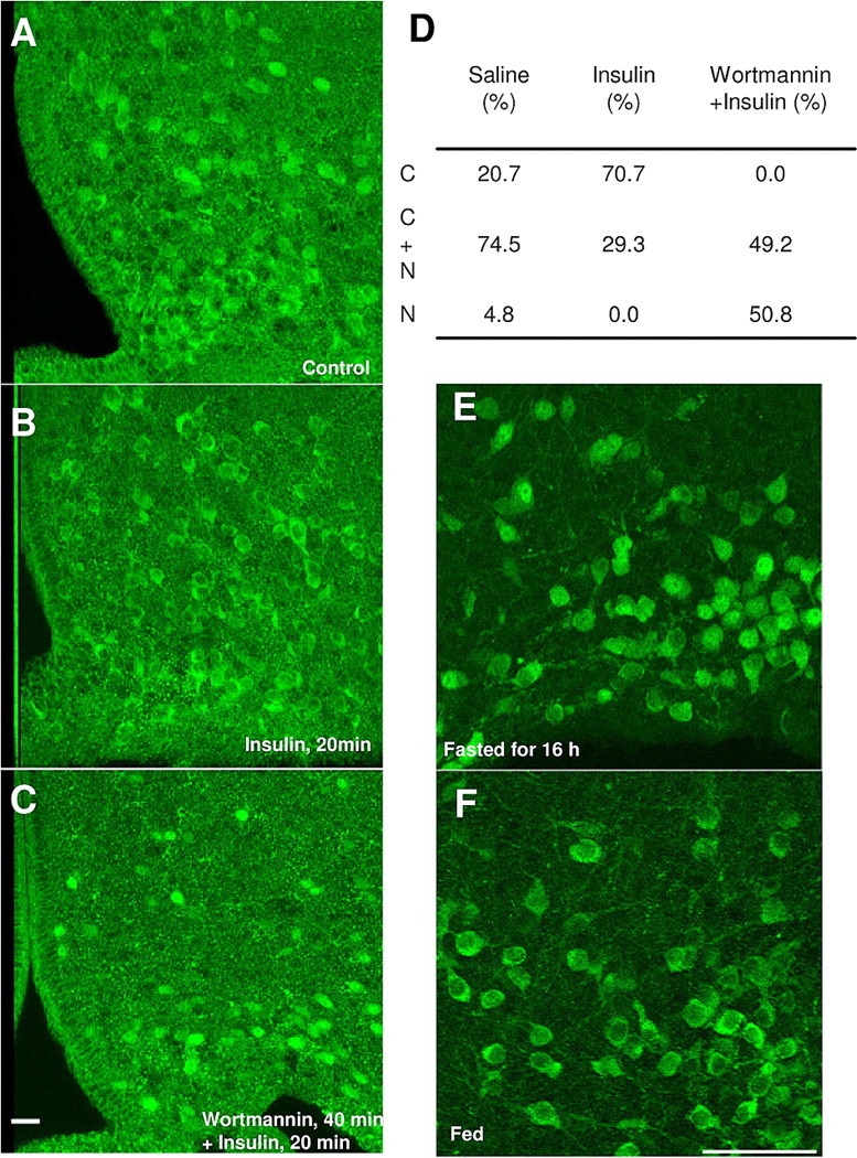
In vivo characterizations of FoxO1GFP mice. A–C, FoxO1GFP-POMC mice fasted for 16 h were administrated with saline (1 μl; i.c.v.) (A), insulin (100 pmol), or wortmannin (100 pmol) (B) 30 min before insulin (100 pmol) (C). Twenty minutes after injection, the mice were perfused and sliced (30 μm). Sections were immunohistochemically stained using an anti-GFP antibody. Scale bar, 20 μm. D, Quantification of FoxO1GFP nuclear translocation in control, insulin or wortmannin plus insulin treated FoxO1GFP-POMC mice. Subcellular localization of FoxO1GFP is presented as the percentage of POMC neurons with C, C+N or N. E, F, Subcellular localization of FoxO1GFP in POMC neurons in FoxO1GFP-POMC mice fasted for 16 h (E) or fed normally (F). Scale bar, 50 μm.