
Figure 3.
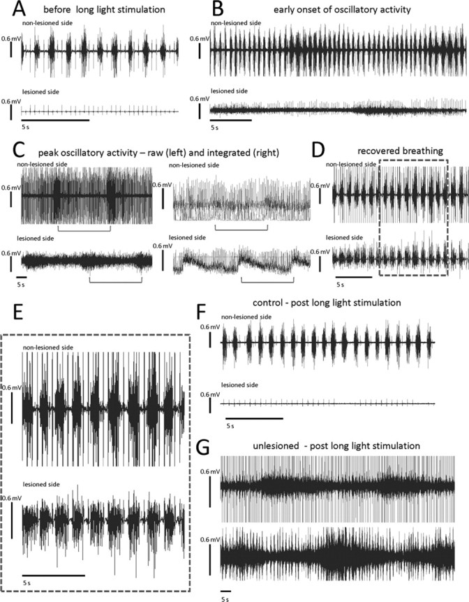
Intermittent photostimulation of ChR2-expressing spinal neurons leads to a pattern of EMG hemidiaphragmatic activity that is close to normal in C2-hemisected animals. A, Before photostimulation, there is no EMG activity ipsilateral to the lesion (bottom trace). Contralateral to the lesion, there is rhythmic EMG respiratory activity (top trace). B, In the same animal, during the photostimulation protocol of 5 min off, 5 min 0.5 Hz stimulation, a trace amount of EMG activity begins to develop ipsilateral to the lesion (lower trace). As the EMG activity begins to dwindle, the contralateral, intact side begins to display an increase of EMG activity (upper trace). C, This cycling of high intensity activity that wanes, while the contralateral side increases activity, continues with each period of high intensity activity being slightly more than the last (C compared with B), and this is after the last round of photostimulation. The left two traces are of the raw EMG signal, and the right is of the same time point but integrated and rectified. Brackets under traces indicate periods between onsets of increased diaphragmatic EMG activity. D, E, Eventually EMG activity becomes closer to normal patterned respiratory EMG activity. E, Inset of D. F, A trace of control-treated animal after photostimulation. G, A representative trace of the waxing and waning exhibited by non-C2-hemisected animals that expressed ChR2 and were photostimulated. Top trace is of the injected side.