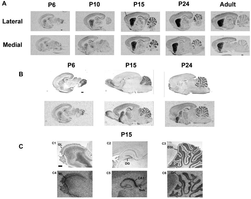
Figure 3.
Rhes mRNA expression in the neonatal period determined by in situ hybridization, sagittal view. Rat pups were sacrificed for analysis on postnatal days 6, 10, 15 and 24, and as adults. A, Medial and lateral sections from rats of the stated ages showing the medial-to-lateral gradient of rhes mRNA in CPu. Hippocampal and cerebellar signal is higher in neonates than in adults. B, Cresyl violet stained sections (top panel) delineate layers of rhes mRNA expression in the same sections (bottom panel). C, higher magnification of P15 from panel B. C1-C3 are cresyl violet-stained sections; C4-C6 show rhes mRNA in the same sections. scale bar = 1 mm (A, B) or 500 μM (C). GL = glomerular layer; DG = dentate gyrus; EGL = external germinal layer; ML = mitral cell layer; Sub = subiculum; GrL = granular cell layer.

