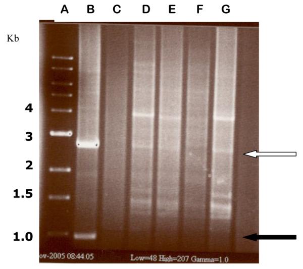
Figure 5.

BamH1-HindIII restriction analysis of representative ampicillin-resistant E. coli transconjugant colonies containing N3::pUC1.0 and R64::pUC1.0 plasmid cointegrates. The DNA’s were analyzed with a rapid boiling DNA extraction and restriction screening method (Epicentre; Madison, WI). Lane A - 1 kb control ladder; Lane B - pUC1.0 control with the 2.9 kb band from pUC19 (white arrow) and the 1.0 kb fragment derived from pSJ5.2 (solid black arrow); Lane C - digested R64::pSJ5.2 DNA; D and E - R64::pUC1.0 transconjugants; Lanes F and G -N3::pUC1.0 transconjugants (the 2.9 kb band from pUC19 is barely visible in lane F). The 1.0 kb BamH1-HindIII fragment is not visible in the cointegrates analyzed from lanes D to G, confirming that integration occurred within that fragment.
