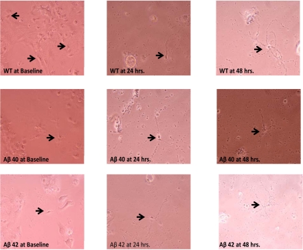
Figure 6.
Cortical neuronal cultures (unstained) at various times following TFW challenge. Conditioned 1-week-old cortical neuronal cultures form 1-day mouse pups were exposed to TFW. Phase contrast images at various time points were obtained (Baseline or 0 hr, 24 hrs and 48 hrs). There was a progressive evolution of apoptotic morphology in cultured cells. This was most marked in the transgenic population (Aβ40 and Aβ42) and somewhat less so in WT. At 24 hours in particular (middle column of images), WT cells exhibited fewer or less robust apoptotic features than either Aβ40 or Aβ42 cells. At 48 hours post challenge, all three populations had become grossly apoptotic.

