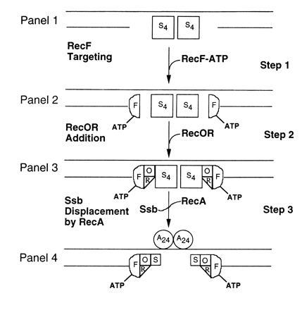
Figure 7.

Cartoon for the RecF–RecO–RecR action in gap repair. For the sake of simplicity only presynaptic events are shown. The first letter of each protein is used to represent the respective protein: S, Ssb; F, RecF; O, RecO; R, RecR; A, 24. (Step 1) gDNA bound by Ssb tetramers (S4). (Step 2) RecF–ATP complex bound at gap junctions of gDNA. It is assumed that RecF protein, in the presence of ATP (step 3) recognizes and subsequently binds at gap junctions. (Step 3) Multiprotein–DNA complexes consisting of RecF–ATP, RecO, RecR, and Ssb. The RecO–RecR are targeted to RecF–ATP–gDNA complexes (step 3) and RecO makes direct contacts with Ssb. (Step 4) Presynaptic filaments consisting of RecF–RecO–RecR–Ssb and RecA proteins. Ssb tetramer displacement is initiated by RecA protein in the presence of RecF–RecO–RecR complex (step 3). Salient features of the cartoon are described in the text.
