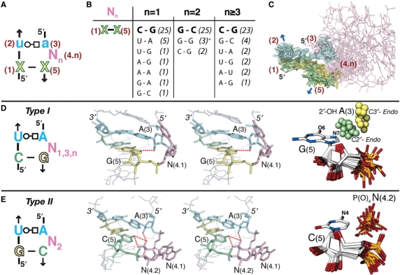
Figure 1.
Type I and type II UA_handle signatures and conformers. (A) The UA_handle is typically formed of a U(2):A(3) (WC:HG) trans bp (in blue) stacked on a classic WC bp, X(1):X(5) [purine in green, pyrimidine in yellow), with one or more bulging nucleotides located 3′ of A(3), (N(4.n) in pink]. For bp annotations, see legend of Figure 2. (B) Sequence variation of X(1):X(5) in function of N(4.n). The table is based on the sequence analysis of 99 atomic structures of UA_h with canonical U(2):A(3) (WC:HG) trans bp. The occurrence of each bp is indicated between brackets. (+) These three G(1):G(5) bps are all from one of the UA_hs of the double T-loop motif. Despite a 2nt bulge, this non-canonical UA_h is of type I. (C) Superposition of canonical UA_handle motifs identified in X-ray structures and listed Table S1. The color code corresponds to the one in (A). The conserved atomic positions in X(1):X(5) and U(2):A(3) were used for the superposition. (D) Sequence signature of type I UA_h: an H-bond can be formed between N7-G(5) and 2′OH-A(3) when the sugar pucker of A(3) is in C2′ endo. (E) Sequence signature of type II UA_h: an H-bond can form between N4-C(5) and the O1 or O2 of P(O)4-N(4.2). As seen in the stereographic image, an additional H-bond can potentially form between 2′OH-A(3) and O2P-N(4.2). For more examples of type I UA_h, see also Figure S1 from Supplementary Data.

