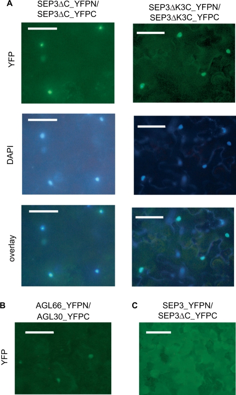
Figure 5.
(A) Subcellular localization and in planta dimerization of SEP3ΔC and SEP3ΔK3C. Proteins are expressed as fusions with the N- (YFPN) or C-terminal (YFPC) part of YFP as indicated above the picture in Nicotiana benthamiana leaves. Leaf sections were analysed with a fluorescence microscope. YFP signals are shown in the upper row, nuclear staining using DAPI is shown in the middle row. The overlay in the lower row shows co-localization of the DAPI and YFP signals. (B) The YFP signals of two proteins known to interact, i.e. AGL30 (fused to the C-terminal part of YFP) and AGL66 (fused to the N-terminal part of YFP) is shown as a positive control. (C) No YFP signal could reliably be detected when full-length SEP3 was tested alone or in combination with a C-terminal deleted version, as exemplarily shown for SEP3 (fused to the N-terminal part of YFP) co-expressed with SEP3ΔC (fused to the C-terminal part of YFP). This might be due to steric hindrance of the C-terminal domain. Scale bars, 100 µm.

