
Figure 1.
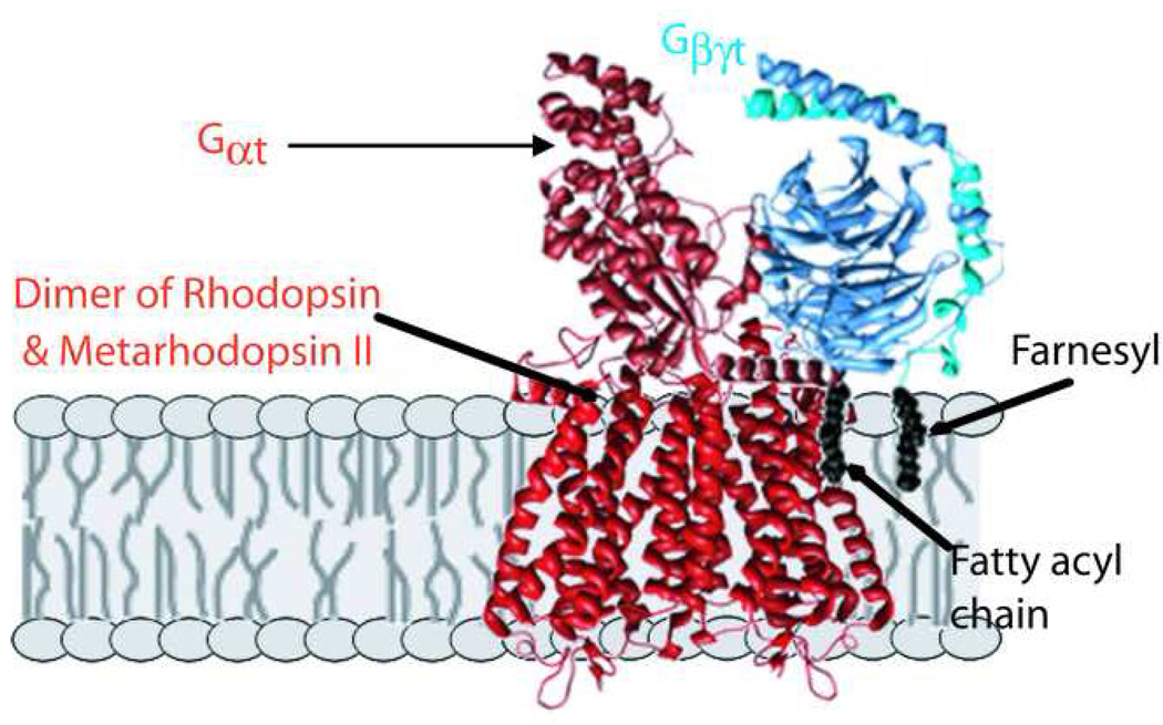
Proposed model for first complex formed in photoactivation. Following absorption of light and photoisomerization by one subunit of the rhodopsin dimer, a heterodimer of rhodopsin and Metarhodopsin II (R*) is formed, which quickly complexes with the G protein, transducin, in its heterotrimeric form. A conformational change within Gαt allows release of bound GDP, which then allows binding of GTP and dissociation of activated Gαt–GTP. The representation of the complex is purely schematic, as only indirect low resolution information is available on the structures of the actual complexes. The major contacts with R* are provided by Gαt, with both its carboxyl and fatty acylated amino termini known to be involved. Gβγ are also important for R* binding, with potential interactions with the membrane hydrocarbon phase provided by the farnesyl group on Gγ. For making the structure images shown, PDB files 1U19.pdb(Okada, et al., 2004) and 1GOT.pdb(Lambright, et al., 1996) were used with UCSF Chimera (Meng, et al., 2006, Pettersen, et al., 2004).