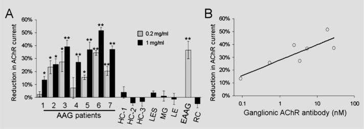
Figure 2. Summary of IgG effect on ganglionic acetylcholine receptor (AChR) current.
(A) Reduction in peak current after 20-minute exposure to IgG is shown for all subjects. IgG from all seven autoimmune autonomic ganglionopathy (AAG) patients and from rabbits immunized against the ganglionic AChR (experimental AAG [EAAG]) produced a current reduction (*p < 0.05, **p < 0.0001 compared with control IgG). IgG from healthy control (HC), Lambert–Eaton syndrome (LES), MG, autoimmune limbic encephalitis (LE), or adjuvant-control rabbits (RC) had no effect on the ganglionic AChR current. Error bars represent SEMs (number of cells for each data point is 3 to 8). (B) The magnitude of current reduction for each AAG patient IgG (at 1 mg/mL) correlated with the serum ganglionic AChR antibody level (log-linear regression, r2 = 0.73, p = 0.01).

