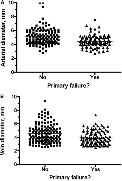
Figure 2.
Scatter plot of the distribution of the (A) arterial diameters and (B) venous diameters of vessels used to create brachiocephalic fistulas. Although the mean arterial and venous diameters were significantly lower in fistulas with a primary failure than in those successfully used for dialysis, there was substantial overlap in the values between the two patient groups.

