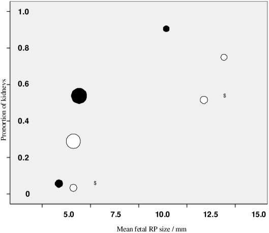
Figure 5.
Proportion of kidneys diagnosed with renal pelvis dilation (RPD) by 6 wk of age. Unfilled circles represent routine studies, and filled (black) circles represent cohorts that included referred patients. Area of circles is proportional to group sample size for all the kidney data. $ represents subgroups within the same study.

