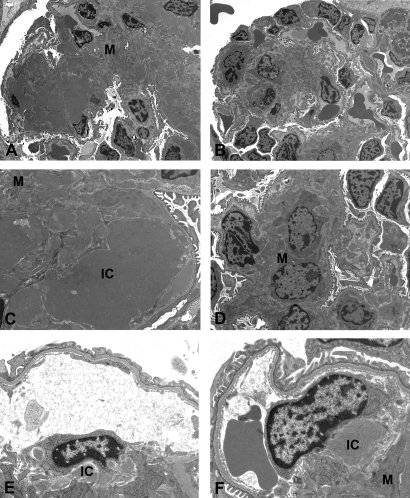
Figure 4.
Electron microscopic features of the glomerular lesions. (A through D) Whereas glomeruli from the vehicle-treated TSLP-Tg mice showed extracellular matrix expansion with massive electron-dense IC deposits in the mesangium as well as in the subendothelial space (A and C), 4 wk of treatment with imatinib results in dramatic reduction of an extracellular matrix expansion and IC deposits (B and D). (E and F) Mice that received imatinib treatment beginning at day 90 also showed a reduction of IC deposition, and macrophages can be seen apparently internalizing these complexes. M, mesangium. Magnifications: ×2400 in A and B; ×7100 in C; ×4400 in D; ×10,400 in E and F.

