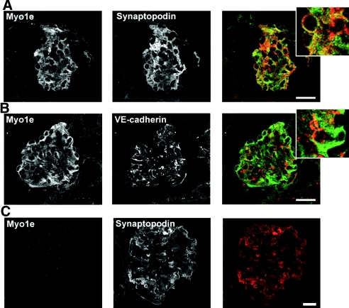
Figure 1.
Myo1e localization in mouse kidney. (A through C) Frozen kidney sections prepared from WT mice (A and B) and Myo1e-KO mice (C) were double labeled with antibodies against myo1e and either podocyte marker synaptopodin or endothelial cell marker VE-cadherin. Myo1e co-localized with synaptopodin (A) but not with VE-cadherin (B). No labeling with Myo1e antibodies was observed in the KO mice (C). Merged images show Myo1e in green and synaptopodin or VE-cadherin in red. Insets in A and B show enlargements of selected regions of the merged images. Bar = 20 μm.

