Figure 5.
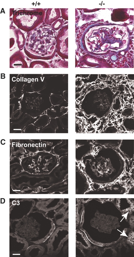
Myo1e-KO mice develop renal inflammation and fibrosis. (A through D) Paraffin-embedded sections (A) or frozen sections (B through D) or of mouse kidneys were stained using Trichrome stain to reveal extracellular matrix deposits (A) or using antibodies against collagen V (B) and fibronectin (C) or mouse C3 (D). (A) Trichrome staining of kidneys from Myo1e-KO mice revealed juxtaglomerular deposition of collagen (blue) and cell infiltration into the interstitial space. (B and C) Immunofluorescence staining indicated increased deposition of collagen V (B) and fibronectin (C) in the kidneys of KO mice. (D) Proximal tubules of Myo1e-KO mice contained granular deposits of C3 (arrows). Complement deposits were not observed in the kidneys of WT mice. Bars = 20 μm.

