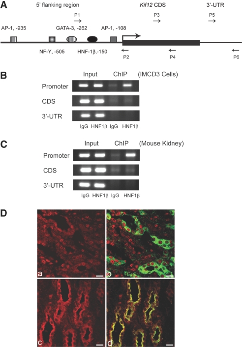
Figure 3.
Validation of the in vivo association of HNF-1β with the Kif12 promoter. (A) Schematic diagram of the mouse Kif12 promoter. Arrows indicate primers that were used for ChIP assays of the promoter (P1 and P2), coding sequence (P3 and P4), and 3′-untranslated region (P5 and P6). (B) Occupancy of the Kif12 promoter by endogenous HNF-1β in chromatin from mIMCD3 cells was verified by ChIP assay. (C) In vivo association of HNF-1β and the Kif12 promoter in chromatin from mouse kidney was confirmed by ChIP assay. (D) Kidney sections from adult mice were co-stained with antibodies against HNF-1β (red, a and b) or Kif12 (red, c and d) and DBA (green, b and d). HNF-1β was localized in the nuclei and Kif12 was localized in the cytosol of DBA-positive collecting duct cells. Bars = 10 μm.

