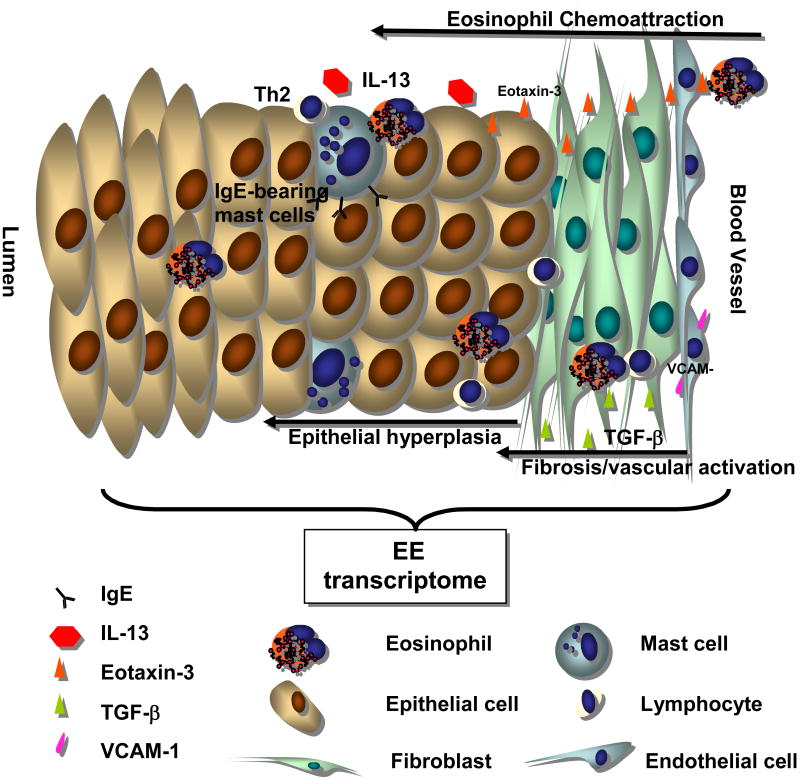
Figure 1. Molecular Pathogenesis of Eosinophilic Esophagitis.
EE arises from the interaction between genetic factors and environmental exposures. This interaction leads to the secretion of a complex array of cellular mediators. The expression of IL-13 and TGF-β likely contributes to the release of eotaxin-3, increased collagen production (fibrosis) and vascular activation (VCAM-1). Finally, IgE can be detected on the surface of mast cells and likely contributes to mast cell activation. This figure was adapted with the publisher's permission.46

