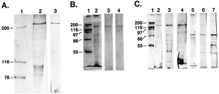
Figure 3.

SDS/PAGE and Western blot analyses of partially purified recombinant fusion proteins of hFAS and its subdomains. (A) Protein standards (lane 1) and the MBP-hFAS fusion protein (5 μg) were subjected to SDS/PAGE analysis (4–15% gradient), and the gel was then stained with Coomassie blue (lane 2) or subjected to Western blot analysis using anti-hFAS antibodies (lane 3). (B) and (C) Protein standards (lanes 1) and 5 μg each of fusion proteins MBP-hFAS-dI (B, lane 2), TRX-hFAS-dII-III (C, lanes 2, 3, and 4), and TRX-hFAS-dII′-III (C, lanes 5, 6, and 7) were subjected to 7.5% SDS/PAGE analysis, and the gels were either stained with Coomassie Blue (B, lanes 1 and 2; C, lanes 1, 2, and 5) or subjected to Western blot analysis using either anti-hFAS antibodies (B, lane 3; C, lanes 3 and 6), anti-MBP serum (B, lane 4), or S-protein AP conjugate (C, lanes 4 and 7).
