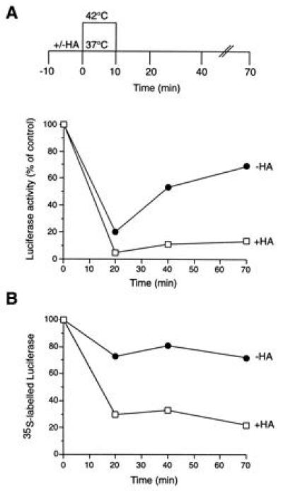
Figure 1.

Effect of heat shock on firefly luciferase expressed in vivo in the presence of HA. Luciferase activities (A) and levels of 35S-labeled luciferase protein (B) in SW620 colon carcinoma cells subjected to heat shock at 42°C and recovery at 37°C in the presence (□) and absence (•) of HA. Ten minutes before heat shock, cultures received 40 μg/ml cycloheximide. Values measured in DMSO-treated control cells maintained at 37°C throughout are set to 100%. Note that, although normally localized in peroxisomes, more than 90% of luciferase expressed in SW620 cells was localized in the soluble supernatant fraction of a high-speed centrifugation (18), as uptake into peroxisomes is inefficient. Luciferase did not form insoluble aggregates during incubation at 42°C.
