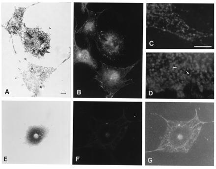
Figure 3.

Bright-field (showing melanosomal distribution) (A and E) and immunofluorescence (B–D, F, and G) photomicrographs of wild-type (A–D) and dv/dv (E–G) primary melanocytes. Antiserum to a dilute tail peptide was used as the primary antibody in all panels except D, for which an antibody to the carboxyl terminus of tyrosinase (αPEP7) was used. G is an underexposure of the same negative shown in F; it shows the background labeling and the extent of the cell’s cytoplasm. The cell at the middle of the left margin of B is a fibroblast that cannot be seen in A. (Bars: A, 10 μm; C, 5 μM. C and D have the same magnification; the remaining panels have the same magnification as A.) The difference between the punctate staining of the anti-dilute antibody and the ring-like staining of the melanosome periphery by αPEP7 (arrowheads) can be seen in at higher magnification in C and D.
