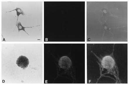
Figure 4.

Bright-field (showing melanosomal distribution) (A and D) and immunofluorescence (B–F) photomicrographs of dilute suppressor (a/a dv/dv dsu/dsu) (A–C) and leaden (C57BL/6J fz ln/ln) (D–F) primary melanocytes, using the anti-dilute tail peptide antiserum as primary antibody. C and F are underexposures of the same negatives shown in B and E, respectively, to show the extent of the cytoplasm of the cells. (Bar = 10 μm.)
