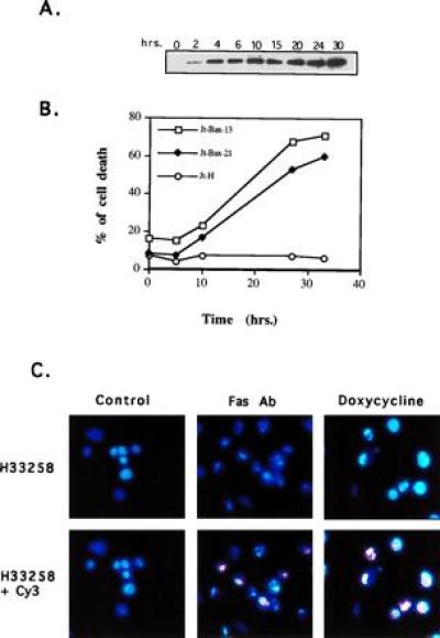
Figure 1.

BAX-induced apoptosis in Jt-Bax cells. (A) Jt-Bax-21 cells were incubated with doxycycline (Sigma) at 1 μg/ml for various times. BAX expression was examined by immunoblot analysis with anti-BAX antibody 651. BAX levels were not substantially greater than that obtained in stable clones. (B) Cell viability was assayed by propidium iodide exclusion. Jt-Bax-13 and -21 were two independent Jt-Bax clones, while Jt-H was an empty vector containing control clone. (C) Jt-Bax-21 cells were treated with anti-FAS antibody (100 ng/ml, Upstate Biotechnology) for 4 hr or doxycycline (1 μg/ml) for 20 hr or left untreated (control). Apoptotic nuclei were visualized by H33258 or H33258 plus dUTP-cyanine-3-TdT-TUNEL (Cy3). The cells with blue or blue plus pink fragmented and condensed nuclei represent apoptotic cells.
