Figure 1.

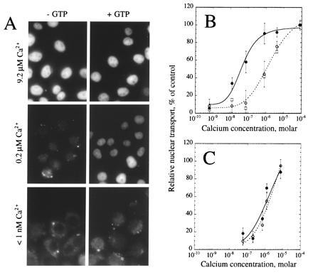
Calcium stimulation of nuclear transport in vitro. Nuclear transport in digitonin-permeabilized HeLa cells was assayed in the presence of a partially fractionated rabbit reticulocyte lysate. (A) Cells were assayed in the absence (Left) or presence (Right) of 1 mM GTP at buffered final free calcium concentrations of 9.2 μM (Top), 0.2 μM (Middle), or less than 1 nM (Bottom). (B) Relative nuclear import was quantified and normalized to control incubations containing 1 mM GTP and 79 μM calcium chloride. Assays were performed in the absence (○, broken line) or presence (•, solid line) of 1 mM GTP, or in the presence of 5 mM GTPγS (□). The data represent the average ± standard deviation (SD) of five (○ and •) or two (□; error bars not included on graph) independent experiments. (C) Prior to assay for nuclear import, thapsigargin was added to the medium on the cells to a final concentration of 10 μM and the cells were incubated for 10 min at 37°C. Thapsigargin was included in all subsequent incubations and washes at a final concentration of 10 μM. Cells were assayed for their ability to support nuclear import in the absence (○, broken line) or presence (•, solid line) of 1 mM GTP at varying calcium concentrations. The data represent the average ± SD of two independent experiments.
