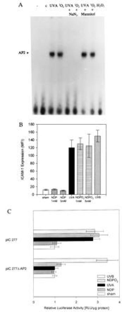
Figure 3.

(A) GEMSAs of nuclear extracts from cultured human keratinocytes. Human keratinocytes were sham-irradiated (C) or stimulated with UVA irradiation (30 J/cm2), NDPO2 (1 mM for 60 min), H2O2 (250 mM) in the presence or absence of sodium azide (50 mM), mannitol (100 mM). Nuclear extracts were prepared 2 h after stimulation as described. Data represent one of two essentially identical experiments. (B) Fluorescence-activated cell sorter analysis of ICAM-1 surface expression in cultured human keratinocytes. Cells were either sham-irradiated, irradiated with 30 J/cm2 UVA radiation (UVA), or incubated in the presence of NDPO2 or NDP at a concentration of 1 or 5 mM for 60 min or irradiated with 100 J/m2 UVB radiation. Cells were harvested after 24 h and were analyzed for ICAM-1 surface expression as described. Data are given as mean fluorescence intensity (mean ± SD of four experiments). C. ICAM-1 promoter activation in 293 cells; 293 cells were transiently transfected with the indicated ICAM-1-based promoter constructs, and ICAM-1 promoter activation was determined as described. Cells were either sham-irradiated (open bars), incubated in the presence of NDPO2 (horizontally striped bars) or NDP (gray bars) at a concentration of 1 mM for 60 min, or exposed to 30 J/cm2 of UVA radiation (solid bars) or irradiated with 100 J/m2 of UVB radiation (diagonally striped bars). Data are given as mean ± SD of relative specific luciferase activity based on total protein contents and represent four experiments.
