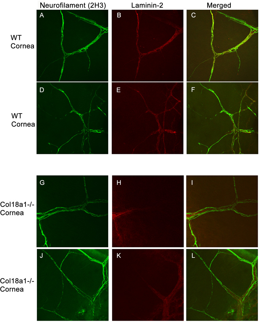
Figure 3. Abnormal laminin-2 staining in the cornea of col18a1−/−.
The cornea was whole-mount immunostained with anti-neurofilament antibody 2H3 (A, D, G, J) and anti-laminin-2 antibodies (B, E, H, K; merged images C, F, I, L, respectively). The corneal limbus is located on the left side of each picture and the central cornea is located on the right. Little to no laminin-2 colocalized with neurofilament in the col18a1−/− corneas (H, K I, L) relative to the WT corneas (B, E, C, F). Laminin-2 staining was only observed at the limbal area of col18a1−/− corneas (H, K), when compared with the WT cornea (B, E).

