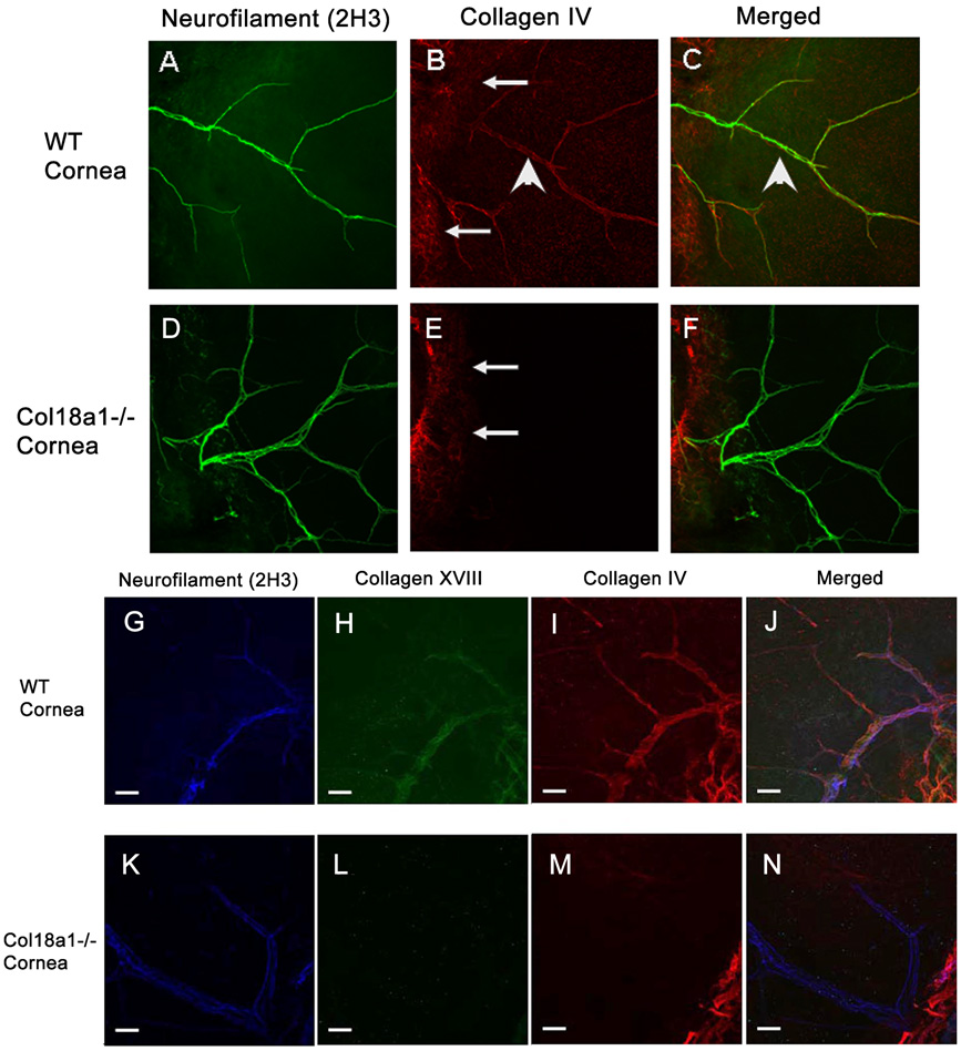
Figure 4. Abnormal composition of the corneal nerve basement membrane in col18a1−/−.
The whole-mount cornea was coimmunostained with anti-neurofilament (A, D) and anti-collagen IV antibodies (merged images C, F, respectively). Almost no collagen IV associated with neurofilament was detected in the col18a1−/− corneas (E) when compared with that of the WT corneas (B). The cornea was then triple stained and visualized using a confocal microscope [antibodies against collagen XVIII (H, L); neurofilament (G, K); collagen IV (I, M); merged (J, N)]. Collagen IV, collagen XVIII and neurofilament colocalized to the nerve basement membrane in WT mice (J). Triple immunostaining did not reveal colocalization of these three molecules in the col18a1−/− mouse corneas (N). The arrowheads point to neurofilament. The arrows point to stromal collagen IV that does not colocalize with the neurofilament.

