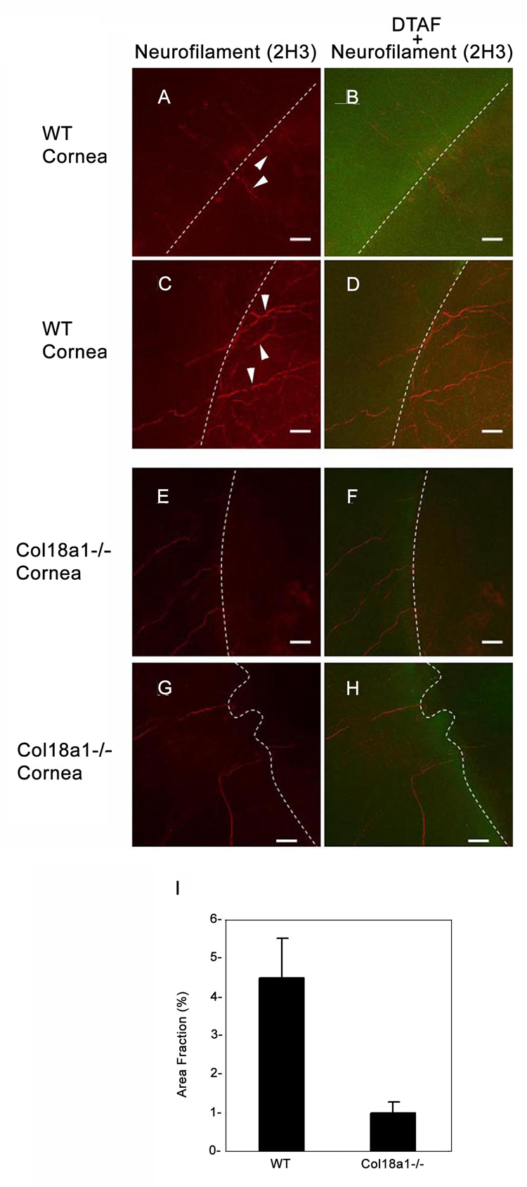
Figure 5. Corneal reinnervation (day 28) after keratectomy was delayed in col18a1−/− mice.
The cornea wound edge was stained with DTAF (green; B, D, F and H) to allow a better comparison with the unwounded area in the peripheral cornea. White dotted lines represent the boundary of the wound edge between the DTAF-stained area and dark DTA-Funstained areas. Regenerating nerves (arrowheads), stained by anti-neurofilament antibodies (red; A and C) passed across the wound edge in WT mice. Regenerating nerves displayed delayed neurite extension into the wounded areas in col18a1−/− mouse corneas (E and G). Semi-quantitative analysis of neurite extension into the wounded corneal area was performed using the Image-J program (I). Four images from WT cornea and four images from the Col18a1−/− cornea were analyzed by adjusting the threshold for accurate representation of the borders of the neurons extending into the injured corneal area. The area fraction of the neurons compared to the entire injured area was calculated and graphed to compare the WT cornea with the col18a1−/− cornea (Bar = 40 µm).

