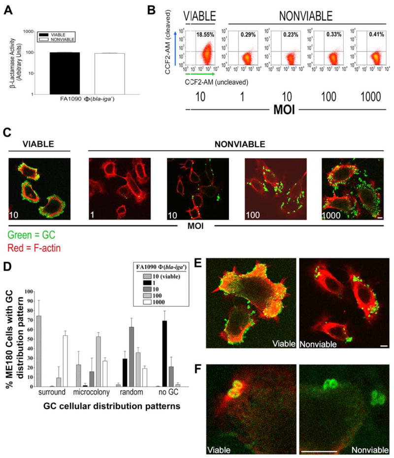
Figure 4.
GC viability is required for host cell F-actin recruitment and subsequent GC internalization by ME180 cells. (A) ME180 cells were incubated with nonviable FA1090Φ(bla-iga′) at different MOI for 6 h and loaded with 0.25 μM CCF2-AM for 45–60 min. ME180 cells incubated with viable FA1090Φ(bla-iga′) at an MOI 10 served as a positive control for internalization/CCF2-AM cleavage. Shown are representative data of five independent experiments. (B) LSCM analyses of the cellular distribution pattern of nonviable FA1090Φ(bla-iga′) at each MOI from part (A). Extracellular GC and F-actin were stained as described in Fig. 2B. (C) Quantification of the GC cellular distribution patterns on approximately 200 randomly selected ME180 cells from the LSCM images in (B) were scored as described in Fig. 2C. Data represents the average (± SD) percentage of ME180 cells that display each distribution pattern from three independent experiments. (D) Shown are higher magnification images of ME180 cells incubated with viable FA1090Φ(bla-iga′) at an MOI 10 (left panel) or nonviable FA1090Φ(bla-iga′) at an MOI 100 (right panel) as in (B). (E) Shown are highly magnified regions of ME180 cells associated with individual viable FA1090Φ(bla-iga′) (left panel) or nonviable FA1090Φ(bla-iga′) (right panel) stained as described in Fig. 2B. Pil+Opa+ FA1090Φ(bla-iga′) were used in all trials. Bars, 5 μm.

