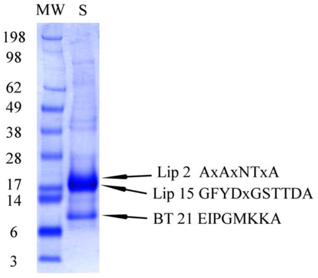
Fig. 7.
1D gel electrophoresis of O. coriaceus salivary gland homogenates. After electrophoresis, the proteins were transferred to a PVDF membrane, stained with Coomassie blue, and bands submitted to Edman degradation. Numbers on the left indicate molecular weight marker positions in the gel. OC-2 and OC-15 are lipocalins; OC-21 is a basic tail protein. For experimental details, see Materials and methods.

