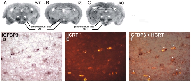
Figure 1. IGFBP3 signals in wild type, ataxin-3 hemizygous, and hypocretin KO mice.
The upper panel shows IGFBP3 ISH staining in wild type (A: WT), ataxin-3 hemizygous (B:HZ) and HCRT knockout (C: KO) mice. HCRT staining in neurons (arrowheads) is markedly reduced or absent in the ataxin-3 mouse. The lower panel shows IGFPB3 ISH signal (D: purple; digoxigenin staining with BCIP/NBT), HCRT fluorescence (E: red; Alexa Fluor) immunostaining, and a composite picture (F), indicating that many hypocretin neurons (asterisks) are positive for IGFBP3 in a WT mouse. Scale bar 20 µm.

