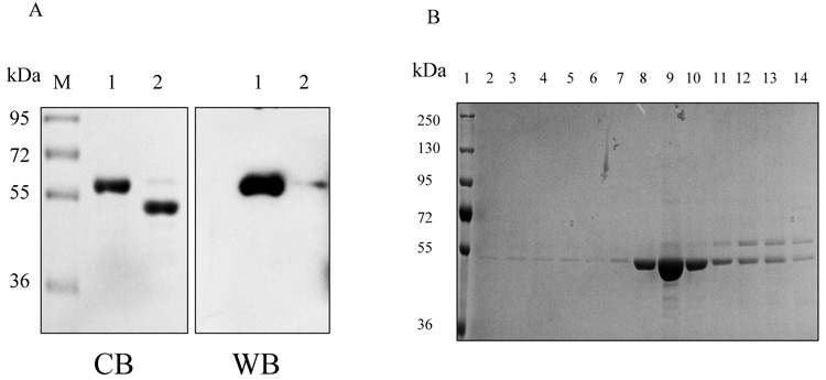
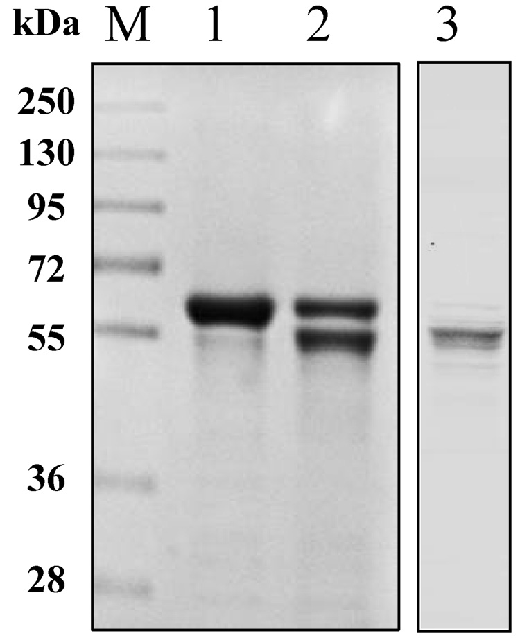
Fig. 3.
Cleavage and removal of His-tag from hALT1 and hALT2 fusion protein. (A) His-tagged ALT1 fusion proteins, undigested (lane 1) and digested (lane 2) by bovine EK, along with molecular mass marker (M), were electrophoresed for Coomassie blue (CB) staining or Western blotting (WB) with anti-His-tag antibody. (B) Coomassie blue staining of EK-digested hALT1 fractions eluted from Ni-NTA agarose column. EK-digested protein mixtures were loaded on to Ni-NTA agarose column, and step-wisely eluted with an increasing gradient of imidazole. Lane 1, molecular mass ladder; lane 2 and 3, flow-through; lane 4~6, column wash; lane7~10, 20 mM imidazole; and lane 11, 12 13 and 14 for 40, 80, 160 and 320 mM imidazole eluents, respectively. (C) Coomassie blue staining of EK-undigested (lane 1), -digested (lane 2), and His-tag free hALT2 (lane 3) eluted from Ni-NTA agarose column at 20 mM imidazole.


