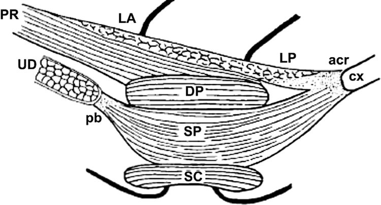
Figure 3.
A sketch of the external anal sphincter from a lateral view, as described by Shafik: External anal sphincter is described as made of 3 loops, basal loop (BL), intermediate loop (IL) and deep loop (DP). Note the relationship between the puborectalis muscle (PR) and DP. We believe that DP is actually the posterior part of the puborectalis muscle (see text for explanation). Adapted from Bogduk N. Issues in Anatomy: the external anal sphincter revisited. Aust N Z J Surg. Sep 1996;66(9):626–629, with permission.

