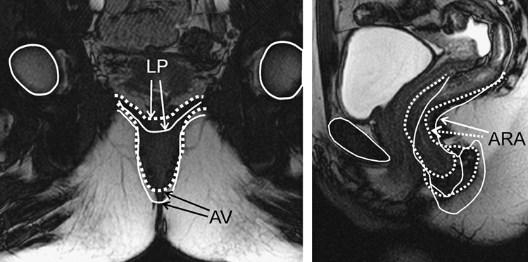
Figure 7.
Magnetic resonance images (MRI) in the mid sagittal and coronal planes: these images were obtained at rest (Solid) and squeeze (dotted) and then the images were overlapped to show the movement of various anatomical structures during squeeze. Note the cranial and ventral movements of the anal canal and change in the anorectal angle with squeeze. In the coronal images, note the vertical movement of the anus, and flattening of the levator plate with squeeze. Obtained from author

