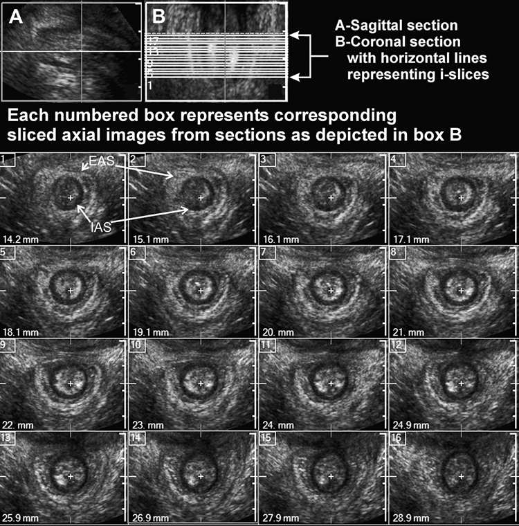
Figure 8.
Ultrasound images of the anal canal obtained from the 3D-US volume: cross-sectional (axial) images along the length of anal canal in a nulliparous subject. In this example the anal sphincter complex is shown at every 1 mm distance using I-Slice function of HD-11 (Philips). Marked in the figure are the IAS (black circle) and the EAS (white outer ring) are smooth, uniform and symmetrical. Obtained from author

