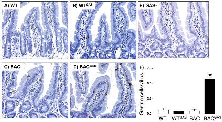
Figure 8. Up-regulation of gastrin cells in the duodenum of gastrin infused hGasBAC mice.
A. Immunohistochemical staining for gastrin in duodenums of A) WT (wild type), gastrin infused B) WT (WTGAS), C) BAC, D) gastrin infused hGasBAC (BACGAS) and E) gastrin-deficient (Gas−/−) mice. Duodenal G cells indicated (arrow). B. Quantification of the number of gastrin positive cells per villus was determined by morphometry. *P<0.05 compared to WT. The results are expressed as the mean ± SEM for 3 mice per group (unpaired t-test).

