
Figure 1.
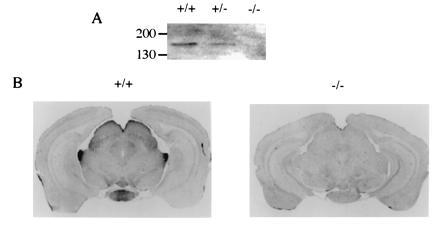
Expression of PLC β4 in visual pathway. (A) Western blot analysis of retinal extracts. Retinal homogenates from a wild-type mouse (+/+) and from mice homozygous (−/−) and heterozygous for disrupted PLC β4 gene were analyzed by Western blotting with the PLC b4-specific antibody. (B) Immunohistochemical staining of brain sections. The wild-type and PLC β4-null mice were perfused with fixative and their brain sections were prepared and stained with a PLC β4-specific antibody using a Vectastain kit (Vector Laboratories).