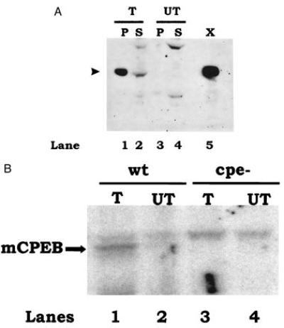
Figure 7.

Recombinant mCPEB recognizes the CPEs of mouse c-mos mRNA. (A) Immunoblot of mCPEB expressed by transient transfection of Cos cells. Cells were transfected with mCPEB cDNA (T) or mock-transfected (UT, no DNA). After an incubation of 36 h, the cells were homogenized and the nuclei were pelleted by centrifugation. Equivalent amounts of pellet (P) and supernatant (S) (approximately one-third of a 100-mm diameter dish) were loaded in a 10% polyacrylamide gel and examined by Western blot analysis using affinity-purified anti-XCPEB antibody. A protein extract corresponding to one-fourth of a Xenopus oocyte (X) was used as a positive control. The size of CPEB is indicated by an arrowhead at the left. (B) UV-crosslinking and immunoselection of mCPEB expressed in Cos cells. Protein extracts from Cos cells that were transfected with mCPEB cDNA (T) or mock-transfected (UT) were UV-crosslinked to radiolabeled c-mos 3′-UTR containing (wt) or lacking (cpe−) CPEs. A supernatant corresponding to 50% of a 100-mm dish was used per lane. Crosslinked products were immunoselected with anti-XCPEB antibody and resolved by SDS/polyacrylamide gel electrophoresis. The results were visualized in a PhosphorImager.
