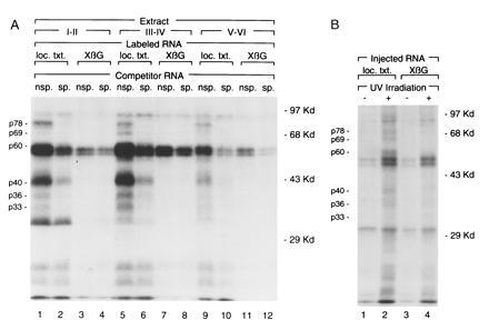
Figure 2.

UV crosslinking of proteins to the localization element. (A) In vitro binding reactions containing S100 extracts, prepared from stage I–II (lanes 1–4), stage III–IV (lanes 5–8), and stage V–VI (lanes 9–12) oocytes, 32P-labeled Vg1 localization transcripts (loc. txt., lanes 1, 2, 5, 6, 9, and 10) or XβG transcripts (lanes 3, 4, 7, 8, 11, and 12) and unlabeled competitor RNA [nonspecific (nsp., lanes 1, 3, 5, 7, 9, and 11) or Vg1 localization transcript (sp., lanes 2, 4, 6, 8, 10, and 12)] were crosslinked by UV irradiation. An autoradiogram of an SDS/10% polyacrylamide gel is shown, with the sizes of molecular weight markers indicated at right and the positions of crosslinked proteins of interest shown at left. (B) 32P-labeled Vg1 localization transcripts (loc. txt., lanes 1 and 2) or XβG transcripts (lanes 3 and 4) were microinjected into stage III–IV oocytes, which were cultured and UV irradiated (+, lanes 2 and 4). An autoradiogram of an SDS/10% polyacrylamide gel is shown. Indicated at right are the sizes of molecular weight markers, and the positions of crosslinked proteins of interest are shown at left.
