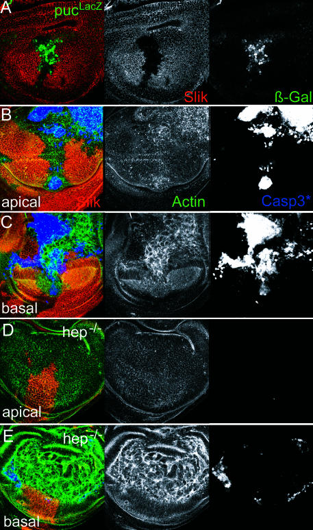
Figure 4. JNK Activity and slik-Dependent Apoptosis.
(A) Wing disc with a Minute+ slik1 mutant clone. Red shows Slik protein. Green shows puc–lacZ reporter gene expression visualized by anti-βGAL. Increased βGAL staining in the clone indicates puc transcription in response to JNK pathway activation.
(B and C) Wing disc with large Minute+ slik1 mutant clones in an otherwise wild-type background. Blue shows activated caspase 3. Green shows actin visualized by phalloidin to show cell outlines. (B) and (C) are different optical sections of the same disc. Genotype: +/Y; FRT42D P(πmyc) M(2)531/FRT42D slik1; hsFLP388/+.
(D and E) Wing disc with large Minute+ slik1 mutant clones in a hemipterous mutant background. (D) and (E) are different optical sections of the same disc. Genotype: hepr75/Y; FRT42D P(πmyc) M(2)531/FRT42D slik1; hsFLP388/+. Note the dramatic increase in clone size, relatively normal apical appearance, and reduction of apoptosis in the clones (detected by activated caspase 3 staining) when JNK pathway activity is reduced in the absence of the hep JNKK.

