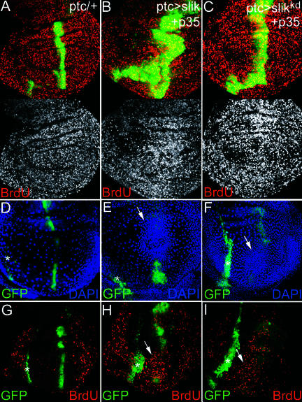
Figure 9. Nonautonomous Stimulation of Cell Proliferation by Slik-Expressing Cells.
(A, D, G) ptcGAL4 UAS-GFP wing discs. (B, E, H) ptcGAL4 UAS-slik UAS-GFP UAS-p35 wing discs. (C, F, I) ptcGAL4 UAS-slikkd UAS-GFP UAS-p35 wing discs. (A–C, G–I) BrdU incorporation (red). (A–C) Projections of several optical sections. (G–I) Sections of the overlying peripodial layer. (D–F) Peripodial cell nuclei visualized by DAPI. Arrows show high nuclear density above the ptcGAL4 UAS-GFP stripe in the columnar epithelium in (E) and (F). Asterisks indicate the peripodial extension of the ptcGAL4 stripe. (H and I) Cells in this region have incorporated more BrdU than control disc in (G).

