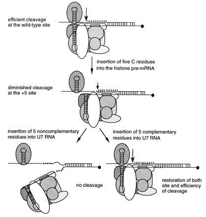
Figure 5.

Models of histone pre-mRNA processing complexes. The wild-type situation is compared with complexes formed on the 5C insertion substrate with the wild-type U7 snRNP, U75nobp snRNP, and the U75bp snRNP. Arrows show the sites of cleavage with levels indicated by arrow size. SLBP is shown as a striped oval, core Sm proteins bound to the Sm binding site (boxed) are different shades of gray, the two known U7 snRNP specific proteins are white ovals, and the hypermethylated cap on the 5′ end of U7 RNA is a solid circle. Base pairing between the U7 RNAs and the substrate HDE sequences is indicated. Insertions into the histone pre-mRNA and the U7 RNAs are illustrated as open and solid circles, respectively. Rigidification of the residues upstream of the HDE is diagrammed as involving protein–backbone contacts (see text).
