
Figure 3.
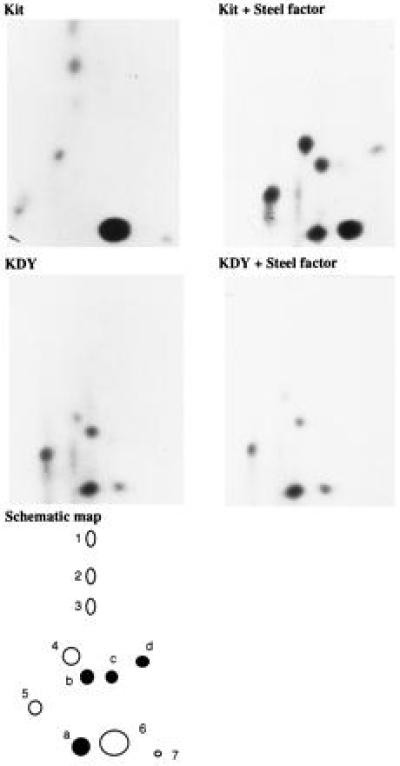
Tryptic phosphopeptide mapping of the Kit and KDY receptors. 32P-labeled Kit and KDY receptors isolated from control and Steel-factor-stimulated cells were digested with trypsin and peptides were resolved by separation in two dimensions on TLC plates. The results are summarized in a schematic phosphopeptide map. The phosphopeptides present in the unstimulated wt Kit receptor, which all contain phosphoserine, are shown as open circles (peptides 1 to 7); phosphopeptides that appear in the wt Kit receptor after stimulation with Steel factor, representing presumptive autophosphorylation sites, are shown as solid circles (peptides a to d).