
FIGURE 3.
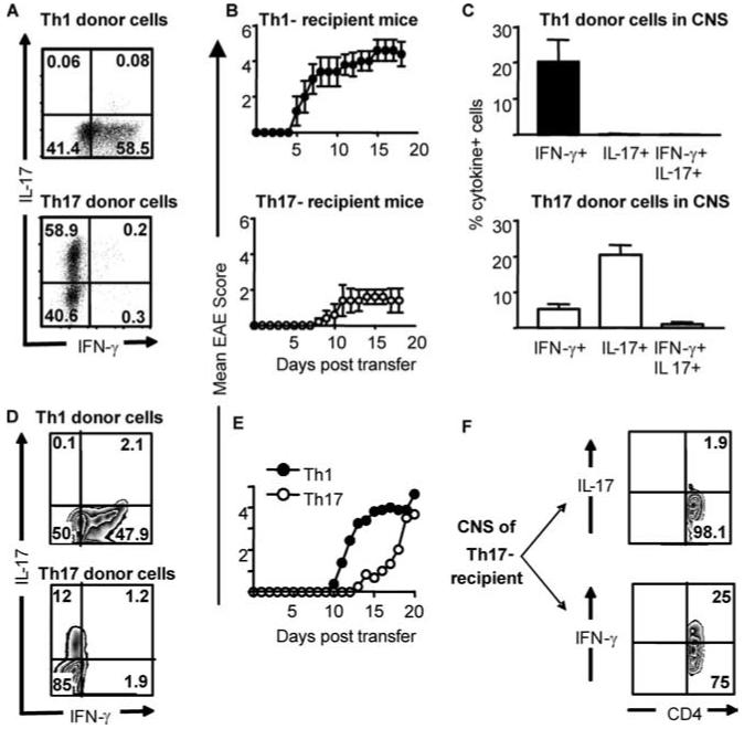
Th17-polarized cells from MBP-reactive TCR transgenic mice can induce mild/delayed EAE, correlating with the in vivo appearance of IFN-γ-producing cells. Th1- and Th17-polarized populations were generated from naive Tg4 mice and transferred into B10.PL mice (A-C) or B10.PL × RAG2-/- mice (D-F). A and D, Intracellular cytokine staining of Th1-polarized (top panels) and Th17-polarized (lower panels) Tg4 cells before transfer. B, Clinical course of EAE induced by the transfer of Th1 (upper panel) and Th17 (lower panel) cells, n = 5. C, Ac1-9 stimulated IFN-γ and IL-17 production by Th1 (upper panel) and Th17 (lower panel) donor cells recovered from the CNS as determined by intracellular cytokine staining. Graphs show the mean ± SD of cells recovered from three surviving Th1-transferred and five Th17-transferred mice. E, Th1 (closed symbols), or Th17-polarized (open symbols) Tg4 T cells (as shown in D) were transferred to B10.PL × RAG-/- recipients (three to four mice per group). F, Ac1-9-stimulated cytokine production by CD4+ T cells recovered from the CNS of a representative B10.PL × RAG2-/- recipient with a disease score of 3. Data are from two of seven experiments giving consistent results.